„SPECTROVERSUM“ - Center for Spectroscopic Characterization of Materials and Electronic/Molecular Processes
Hosted by Vilnius University and Center for Physical Sciences and Technology

PARTNERSHIP WITH MAX IV
 |
 |
RI SPECTROVERSUM and Swedish National Laboratory of Synchrotron Radiation MAXIV (https://www.maxiv.lu.se) in 2019 has signed 8 years collaboration agreement for performing experimental research in broad spectroscopic range. In the frame of this agreement we offer:
- Consultations about possibilities to perform experiments at MAXIV laboratory
- Consultations and assistance in finding appropriate Beamline for your experiments
- Assistance in the preparation of application for the beam-time at MAXIV beamlines
- Assistance in starting scientific collaborations with research groups from SPECTROVERSUM and MAXIV
- Up to 1-month support for PhD’s and postdoc’s to cover travel and living expences in Lund.
We are looking forward to the fruitful work together- Spectroscopy in broad range of electromagnetic radiation of various samples, starting from single molecule and ending with proteins and biological tissues.

Members of SPECTROVERSUM -MAXIV collaboration steering committee at MAXIV are discussing future of the joint scientific projects
Contact: Prof. Valdas Sablinskas - valdas.sablinskas@ff.vu.ltvaldas.sablinskas@ff.vu.lt
SERVICES
1. IR Absorption and Raman Spectroscopy
Contact person – Valdas Šablinskas, valdas.sablinskas@ff.vu.lt
Description of the services
Infrared absorption and Raman spectroscopy are effective methods for material characterization. When light is interacting with a molecular compound, states of molecular vibrations are changing. For this reason, both IR absorption spectroscopy and Raman spectroscopy allow obtaining straightforward information about chemical composition, structure and other features of the material.
These methods are applied for analysis of the following systems and processes:
1. IR spectroscopy of unstable molecular compounds and complexes isolated in inert gas matrices;
2. IR spectroscopy of bio-molecules in solutions and on surfaces;
3. Raman spectroscopy of phonons in crystal structures;
4. IR microspectroscopy of organic compounds in aerosols;
5. IR microspectroscopy of biological tissues;
6. Vibrational (IR and Raman) spectroscopy of dyes in works of art;
7. Vibrational (IR and Raman) spectroscopy of coatings and polishers in odontology;
8. Structural analysis of biomolecules in solution.
9. Hydration studies of biomolecules.
10. Analysis of Raman spectra of semiconductors.
11. Analysis of biomolecular films on surfaces.
12. Qualitative analysis of gemstones (diamond, sapphire, etc.) by means of Raman spectroscopy;
13. Analysis of Raman spectra of ceramics and organic/inorganic crystals.
14. Analysis of carbon nanomaterials, diamond-like coatings and graphene by means of Raman microspectroscopy and RR (resonance Raman) spectroscopy.
2. Non-linear and Multimodal Raman Spectroscopy
Contact person – Gediminas Niaura, gediminas.niaura@ftmc.lt
Description of the services
Raman spectroscopy is effective methods for characterization of material. When light is interacting with the material, states of molecular vibrations are changing. For this reason Raman spectroscopy allows obtaining straightforward information about chemical composition, structure and other features of the material. For Raman studies of low concentration samples various resonance methods such as Resonance Raman and Surface enhanced Raman (SERS) can be applied. The following systems and processes can be studded at SPECTROVERSUM using various Raman approaches:
Raman spectroscopy:
1. Structure of biomolecules in solution.
2. Hydration studies of biomolecules.
3. Analysis of biomolecular films on surfaces.
SERS Spectroscopy:
1. In situ SERS study of adsorption, redox transformations and function of amino acids, peptides, proteins, and other biomolecules.
2. Potential-dependent studies of biomolecules adsorbed on Au, Ag, and Cu electrodes.
3. Interaction of biomolecules with self-assembled monolayers of functionalized thiols.
4. Analysis of self-assembly of biomolecules at surfaces.
5. Investigation of structure and function of hybrid membranes at metal surfaces.
Resonance RAMAN Spectroscopy:
1. Redox transformations of active centers of metalloproteins, estimation of redox potential.
2. Analysis of adsorption of metalloproteins.
3. Interacton of biomolecules with conducting polymers at electrochemical interface.
4. Interaction of biomolecules with graphene and carbon nanotubes.
3. Steady State Fluorescence and UV-VIS Spectroscopy
Contact person – Justinas Čeponkus, justinas.ceponkus@ff.vu.lt
Description of the services
1. Measurement and analysis of electronic transmission spectra of solids and fluids.
2. Measurement and analysis of electronic specular reflection spectra of solids and fluids.
3. Determination of shape and size of metal nanoparticlesin colloidal solutions.
4. Measurement and analysis of fluorescence spectra of solid and liquid molecular compounds in 200-900 nm spectral range with pulsed xenon lamp excitation.
4. Ultrafast Electron Spectroscopy
Contact person – Vidmantas Gulbinas, vidmantas.gulbinas@ftmc.lt
Description of the services
1. Characterization of absorption and fluorescence spectra of condensed materials.
2. Measurement of the fluorescence decay kinetics and characterization of the fluorescence evolution dynamics.
3. Characterization of electronic and conformational relaxation processes in organic molecules, molecular compounds and nanostructures.
4. Characterization photoelectrical properties of organic semiconductors.
5. Characterization of fluorescence properties of single molecules.
5. Mass Spectrometry
Contact person – Andrius Garbaras, andrius.garbaras@ftmc.lt
Description of the services
1. Stable isotope analysis of the size-segregated atmospheric and combustion aerosols.
2. Stable isotope as tracers for the evaluation of tropic position and food webs of the animals.
3. Neutron irradiation reconstruction in the reactor graphite using 13C/12C ratio.
4. Analysis of carbon and nitrogen variations in the sediments of Curonian lagoon and the Baltic Sea.
5. Stoichiometry determination in bionanocomposites.
6. Food authentication (e.g. detection of adulteration of sugar in fruit juice).
7. Ecology (feeding habits, food webs, invasive species etc.).
8. Geology (sedimentation processes, climate reconstruction etc.).
9. Hydrology (water sources, precipitation).
Laboratory is seeking ISO 17025 accreditation on the method ENV 12140 ”Fruit and vegetable juices – Determination of the stable carbon isotope ration (13C/12C) of sugars from fruit juices – Method using isotope ratio mass spectrometry”.
6. NMR and EPR Spectroscopy
Contact person - Vytautas Balevičius, vytautas.balevicius@ff.vu.lt
Description of the services
1. Structure elucidation of organic compounds applying High Resolution 1D/2D NMR techniques.
2. Structure elucidation of organic and inorganic solids applying Solid State Magic Angle Spinning (MAS) techniques.
3. Size profile determination in nano-structured complex solids applying CP-MAS technique.
4. Quality monitoring for chemistry, drugs, dye, food, etc. industry.
5. Structural studies of materials containing transition metal ions such as Fe, Cu, Mn, Co, Mo or Ni.
6. Studies of molecular substances containing free radicals.
7. Studies of defects induced by impurities in semiconductors, ionizing radiation or N centers in diamond.
7. Terahertz and Microwave Spectroscopy
Contact person – Šarūnas Svirskas, sarunas.svirskas@ff.vu.lt
Description of the services
1. Calculation of dielectric permittivity spectrum from complex transmission coefficient;
2. Incorporation of THz dielectric spectra with lower frequency measurements (10 mHz – 100 GHz);
3. Determination of THz spectra in ceramics and crystals;
4. Measurements of molecular vibrations in semi crystalline polymers;
5. Measurements of THz spectra in thin films;
6. Combination of THz and IR spectra for determination of dielectric permittivity.
8. Synthesys of perovskites by solid state reaction
Spectroversum services related to Synthesys of perovskites by solid state reaction
1. Synthesis of perovskite oxides by solid-state reaction method.
2. Production of perovskite piezoelectric layers by coating method;
3. Production of powders with controlled grain size;
Equipment for solid state oxide reaction synthesis
Available equipment:
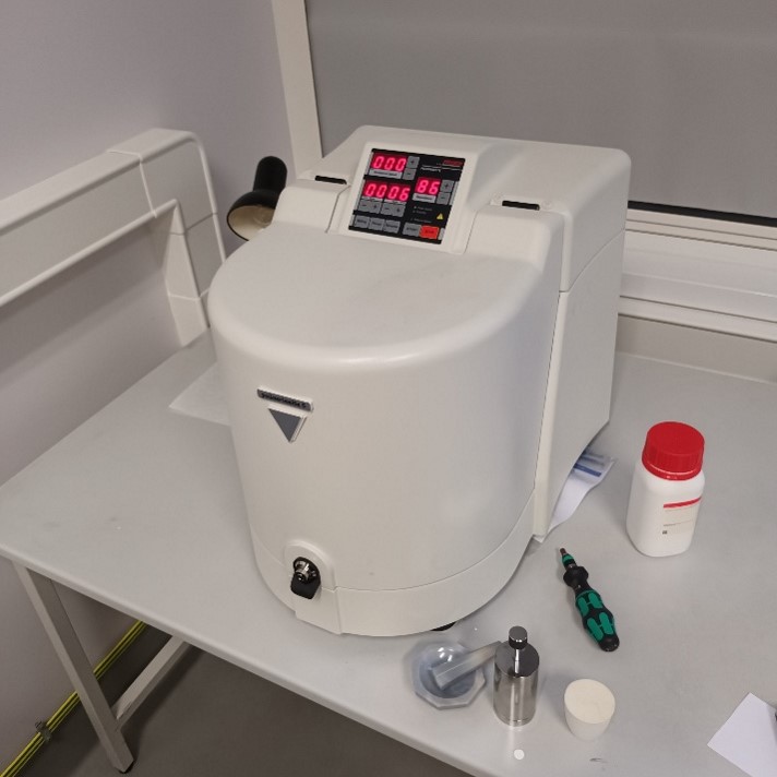
1. Planetary mill;
2. SNOL furnace (maximum temperature - 1250 °C);
3. Polisher/Grinder;
4. Specac hydraulic press (10 t);
5. Moulds (25.4 mm (1 inch); 8 mm; 5 mm);
6. Precision scales.
 |
 |
RESOURCES
IR absorption and Raman spectroscopy
INSTRUMENTATION FOR INFRARED SPECTROSCOPY
1. FTIR spectrometer Vertex coupled with infrared microscope Hyperion 3000 (Bruker).

Spectral range in sample compartment 50-7000 cm-1;
Spectral range in microscope 600-5000 cm-1;
Spectral resolution 0.5 cm-1;
Microscope lateral resolution up to 10 microns;
Sample spectral imaging and mapping with single or focal plane array detectors (64x64 pixels);
Possible measurement modes: transmission, reflection and ATR;
Possibility to use controlled temperature cryostat Linkam, working range 90 – 400 K;
Possibility to use high passlength low pressure gas cell 60 m optical passlength.
2. FTIR spectrometer Alpha (Bruker), modified for Fiber optics based ATR measurements (Artphotonics).

Portable spectrometer with possibility to measure in situ, no need to move the sample to the spectrometer;
Spectral range with standard diamond ATR 350-7000 cm-1;
Spectral range with fiber optics 600-3500 cm-1;
Spectral resolution 1 cm-1.
3. High resolution FTIR spectrometer IFS120 (Bruker).

Spectral range 100-25000 cm-1;
Spectral resolution 0.00125 cm-1;
Coupled with long pass low pressure gas cell optical path up to 100 m;
Coupled with low temperature cryostat lowest temperature 3 K.
INSTRUMENTATION FOR RAMAN SPECTROMETRIC MEASUREMENTS
1. FT-Raman spectrometer MultiRam coupled with Ramscope microscope (Bruker).

Excitation wavelength: 1064 nm (Nd:YAG laser, power – up to 1 W); 785 nm (DPSS laser power up to 500 mW) power adjustment from 0 to 100% with 1% step;
Useful spectral range: 3600 – 50 cm-1 (Raman shift) with 1064 nm excitation; 5000‑50 cm-1 (Raman shift) with 785 nm excitation;
Spectral resolution: ≥0,5 cm-1;
Possibility to control sample temperature in range: -196 °C – 600 °C;
Possibility to investigate small samples down to 100 microns;
Possibility for spectral mapping;
Types of samples - liquid, solid, powder.
2. Confocal Raman Grating Spectrometer with microscope „MonoVista CRS“, S&I GmbH.

Excitation wavelength: 457 nm (DPSS laser power up to 50 mW), 532 nm (DPSS laser power up to 500 mW), 633nm (DPSS laser power up to 30 mW) 785 nm (DPSS laser power up to 100 mW), power adjustment possibility;
Spectral range: 5000 – 60 cm·¹ (Raman shift) with edge filters for all excitation wavelength;
Low frequency measurements down to 5 cm-1 with Bragg filters for 532 nm excitation;
Spectral resolution: ≥1 cm-1;
Microscope and macrochamber for different size samples;
Microscope with 10x, 40x and 100x objectives;
Possibility to control sample temperature in range 90-400° K;
Possibility to investigate small samples down to 100 microns;
Types of samples – gas, liquid, solid, powder.
INSTRUMENTATION FOR LOW TEMPERATURE IR OR RAMAN SAMPLE HANDLING
1. Cryogenic system for vibrational spectroscopy Janis/Sumitomo.

Cryogenic system for low temperature spectroscopy;
Temperature range 3-350 K;
Temperature control accuracy 0.1 K;
Sample solid samples, or sample introduced by condensation from the vapor phase;
Matrix isolation technique;
Adaptable for spectroscopic measurements from 200 – 50 000 cm-1(50μm – 200 nm).
2. Controlled temperature heating-cooling sample stage Linkam FTIR 600.

Temperature range from 77 K to 600 K, temperature control accuracy 0.1 K;
For solid samples;
Adaptable for spectroscopic measurements from 500 – 50 000 cm-1 (20μm – 200 nm).
Non-linear and multimodal Raman spectroscopy of biological objects
Renishaw inVia Raman spectrometer with 7 excitation wavelengths.
 |
 |
Excitation wavelengths:
325 nm (4.25 mW at the sample), low frequency cut-off: 250 cm-1;
442 (52.8 mW), low frequency cut-off: 120 cm-1;
532 (45.3 mW), low frequency cut-off: 70 cm-1;
633 (9.4 mW), low frequency cut-off: 60 cm-1;
785 nm (92 mW), low frequency cut-off: 100 cm-1;
830 nm (166 mW), low frequency cut-off: 100 cm-1;
1064 nm (121.2 mW), low frequency cut-off: 100 cm-1;
“Noch” filter for 532 nm for low frequency (cut of: 10 cm-1) and anti-Stokes measurements, “Noch” filter for 785 nm for low frequency (cut-off: 10 cm-1), “Next” filter for 633 nm for low frequency.
Objectives: VIS 100x/0.85, 50x/0.75, 20x/0.40, and 5x/0.12; Near UV 15x and 40x.
Gratings: 600, 830, 1200, 1800, 2400, 3000 and 3600 lines/mm.
Detectors: UV and NIR enhanced Deep Depletion CCD (1024 x 256 pixels, -70 oC); iDus and InGaAs (512x1 pixels, -90 oC).
Potentiostat: AutoLab PGSTAT101.
Custom equipment: spectroelectrochemical cell, Au, Ag, and Cu electrodes pressed into Teflon (5-6 mm diameter), spectroelectrochemical cell moving mechanism to reduce the laser induces damage to the sample (moving speed 15-25 mm/s).
Vibrational Sum-Frequency Generation Spectrometer (EKSPLA).

|
Schematic layout of a table-top VSFG spectrometer produced by EKSPLA |
Schematics of VSFG generation at the lipid/water interface with adsorbed biomolecule |
We use a commercially available picosecond scanning VSFG system from EKSPLA (UAB, Lithuania). It consists of a Nd:YAG picosecond pump laser and an optical parametric generator (OPG) with a difference frequency generation (DFG) module (Fig. 1). To generate VSFG two ultrashort laser pulses, one of a visible light and another in the infrared region, are overlapped onto the sample surface (Fig. 2). VSFG is enhanced when the infrared is in a resonance with the vibration of a molecule at the surface. Its surface specificity arises due to the selection rule of the undergoing second-order nonlinear process: for such a process the medium must be non-centrosymmetric. VSFG is a very versatile technique that can be used to study many different surfaces and interfaces such as solid/air, solid/liquid, liquid/air; however, the greatest potential of VSFG lies in its ability to measure liquid surfaces with a sensitivity of a few molecular layers.
List of parameters:
Nd:YAG picosecond laser: 1064 nm wavelength, 30 mJ energy, 28 ps pulse duration, 50 Hz repetition rate.
Sum frequency generation range: from 1000 cm-1 to 4000 cm-1.
Energy of infrared beam: 50 – 200 mJ.
Spectral resolution: 6 cm-1.
Wavelength of visible beam: 532 nm.
Energy of visible beam: ~350 mJ.
Incident angles of infrared and visible beams: 55° and 60°.
VSFG system can be equipped with Langmuir-Blodgett Trough (KCV NIMA): surface area – 98 cm2, trough inner dimensions – 195 x 50 x 4 mm, maximum compression ratio – 5.2, barrier speed – 0.1 – 270 mm/min.
Absorption and Steady State Fluorescence spectroscopy
INSTRUMENTATION FOR ABSORPTION MEASUREMENTS IN UV-VIS-NIR- INFRARED REGION
UV-VIS-NIR spectrometer Lambda 1050 (Perkin-Elmer).

Spectral region: for transmission - 175 – 3300 nm, for reflection – 190 – 3300 nm;
Spectral resolution: UV-VIS spectral region - ≥0.05 nm, NIR spectral region – ≥0.20 nm;
Possibility to control temperature in the region - 0 – 100°C , precision - ±0.1°C;
Possibility to change angle of incidence for reflection measurements from 8° to 65°;
Possibility to perform polarization measurements;
Types of samples: gases, liquids, films, monolayers, solids, surfaces.
INSTRUMENTATION FOR FLUORESCENCE MEASUREMENTS
Fluorescence spectrometer for steady state and kinetic measurements Carry Eclipse (Agilent).

Spectral region: 200-900 nm;
Spectral resolution down to 0.1 nm;
Excitation source pulsed Xenon lamp with selectable filters;
Gating times for kinetic measurements from 1μs to 10s;
Automated polarization measurements;
Controlled temperature liquid sample cells 0 – 100°C, precision - ±0.1°C;
Fiber probe.
Ultrafast electronic spectroscopy
1. Spectroscopic complex for characterization of ultrafast electronic processes in condensed materials.

The complex is based on the femtosecond laser, which is used as a source of ultrashort light pulses used for realization of transient absorption, electric field modulated transient absorption, conventional time-of-flight and transient electric field induced second harmonic generation techniques. These techniques allow investigation and characterization of electronic properties of organic materials such as: spectral properties and relaxation times of electronic excitations, charge carrier generation processes and dynamics of the charge carrier mobility in a wide time range.
2. Ultrafast fluorescence spectrometer.

Streak camera is a device used to measure ultra-fast fluorescence. Several of the many advantages against other fluorescence measuring systems is that streak camera can register the entire data-set in very short time and it has very high time resolution – up to 2 ps. Our system uses 3W 1030nm "Pharos" as the main oscilator and "Hiro" harmonics genereator to produce laser pulses of ~80fs and 1030nm, 515nm, 343nm or 257nm, which are used for the sample excitation. Frequency of the main oscilator (76MHz) can be reduced to 10kHz using Pockels cell.
3. Time-correlated single photon counting fluorescence spectrometer.

The FLS920-t is a modular, computer controlled fluorescence lifetime spectrometer.
Based on an L – geometry hardware configuration, the FLS920-t utilises the technique of Time Correlated Single Photon Counting (TCSPC) to measure time resolved luminescence spectra and luminescence lifetimes spanning the range from 100picoseconds to 10microseconds, with the accuracy and the resolution that only the technique of TCSPC can offer.
Features Include:
• Fluorescence lifetimes from 100picoseconds to 10microseconds
• High dynamic range and temporal resolution
• Time Correlated Single Photon Counting operation
• PC plug-in card for TCSPC
• Fully computer controlled through Windows (XP and VISTA 32 bit) software
• Full data reconvolution using a non-linear least square fitting routine
4. CARS and multiphoton fluorescence microscope.

The microscope developed on a base of picosecond laser and inverted microscope is designed for investigation of the microscopic structure of biological objects and other molecular structures with submicrometer spatial resolution. The two nonlinear optical processes, coherent antistokes Raman scattering and nonlinear fluorescence, enable revealing invisible structure of the investigated objects and distribution of some specific molecules.
5. Single molecule spectrometer.

Single-molecule fluorescence spectrometer is for detecting fluorescence spectral dynamics from individual molecules immobilized on a surface or freely diffusing in solution. The setup can be operated in two excitation-detection configurations:
• By exciting a wide sample field in total internal reflection (TIR) mode and detecting fluorescence signal from a number of individual molecules in parallel. Fluorescence image is split into two spectral components which allows observation of FRET in the donor-acceptor dye pair. The main advantage of this detection mode – the possibility of observation of a large number of molecules in parallel. The temporal resolution is limited, however, to approximately a video rate.
• By exciting individual molecules in confocal mode and monitoring fluorescence from separate molecules one at a time. In this mode it is possible to acquire full fluorescence spectra. Temporal resolution for spectral acquisition is a few milliseconds, whereas if opting for the detection of only two spectral components, temporal resolution is up to a few tens of nanoseconds.
Moreover, in confocal mode it is possible to collect fluorescence bursts from molecules diffusing freely in solution. This way vast data statistics are collected relatively fast. By analyzing such data with correlation analysis, it is possible to determine, for example, the efficiency of the excitation energy transfer inside the molecule of interest.
Mass spectroscopy
INSTRUMENTATION FOR MASS SPECTROMETRY
1. Elemental analyser (Flash EA 1112) connected to isotope ratio mass spectrometer (Thermo Delta V Advantgage) for bulk isotope analysis EA-IRMS. Analytical services: carbon (δ13C), nitrogen (δ15N), sulphur (δ34S) stable isotope ratio in the samples (bone collagen, sediments, etc).
 |
 |
2. Gas delivery system Thermo Gasbench II, ) connected to isotope ratio mass spectrometer (Thermo Delta V Advantgage). Analytical services: Oxygen (δ18O), carbon (δ13C) stable isotope ratio in the carbonates, δ2H in water.

3. Gas chromatograph (Thermo Trace GC) ) connected to isotope ratio mass spectrometer (Thermo Delta XP Advantgage) for compound specific analysis GC/C/IRMS. Analytical services: amino and fatty acids nitrogen and carbon isotope ratio in individual compounds, PAH analysis, etc.

4. Gas chromatograph Thermo Trace 1300 connected to mass spectrometer Thermo ISQ7000 for GC-MS analysis. Analytical services: quantification of amino and fatty acids, PAH, etc. Detection limit 10-9.

5. Inductively coupled plasma mass spectrometer Thermo Element 2 for ICP-MS analysis. Analytical services: elemental and isotopic composition of the sample. Detection limit 10-12.

6. Accelerated mass spectrometer (SSAMS, National Enlectrostatics Corporation, USA), with graphitisation system (AGE3, IonPlus, Switzerland) and gas delivery sistem for radiocarbon analysis. Analytical services: radiocarbon age determination, (14C amount in the sample). Needed amoun for the analysis – 1 mg.
 |
 |
NMR and EPR spectroscopy
INSTRUMENTATION FOR NMR SPECTROMETRIC MEASUREMENTS
NMR spectral experiments are performed with 400 and 600 MHz wide and standard bore Bruker spectrometers.

a) High resolution solid state NMR experiments are performed using CP/MAS: triple resonance HXY probe; magic angle spinning (MAS) rate – up to 35 kHz; tuning range of the X channel for predefined nuclei in the range 31P to 13C; tuning range of the Y channel from 23Na to 15N, high-power proton decoupling; temperature range from - 140 °C to +150 °C.
b) Solid state broad band experiments: tuning range - from 109Ag to 31P. Temperature range: from -150 °C to 400 °C.
c) Low temperature solid state experiments. Tuning range from 109Ag to 31P. Temperature range 8-300 K.
d) High resolution liquid state experiments: 1H optimized triple resonance probe designed for 1H observation and 1H observation with 13C/BB decoupling.
INSTRUMENTATION FOR EPR SPECTROMETRIC MEASUREMENTS
The measurements are performed with EPR spectrometer ELEXSYS 560 (Bruker).

Continuous wave (CW) and pulse (FT) modes
Temperature range: 3.8 – 300 K;
Programmable one axe goniometry. Precision - 1/8 degree;
International standard of sensitivity: S/N 2500:1;
Absolute sensitivity: 1.2 x 10⁹ spin/G;
Operation in FT mode
Sensitivity: S/N 200:1 per 10 s, using 10 μM TEMPOL toluene solution;
Channels of strong pulses: four with 1 kW power each;
Time resolution of pulses: 2 ns;
Digital processing of the signals: FFT, high speed 1D and 2D experiments.
Pulsed ENDOR unit
Pulse EPR/ENDOR probes for X and Q band frequencies;
150 W radiofrequency amplifier, the highest achievable radio frequency 250 MHz; Pulse forming unit with two additional microwave pulse excitation pathways in the microwave bridge with the ability to independently change the amplitude and phase.
Terahertz and FIR spectroscopy
INSTRUMENTATION FOR DIELECTRIC (IMPEDANCE) SPECTROSCOPY
Temperature range: 30 – 800 K.
Solartron ModuLAB XM MTS system

Frequency range: 10 µHz – 1 MHz;
Max voltage (AC +DC): ±100 V;
Max current: ± 2 A
Current resolution: 0.15 fA
INSTRUMENTATION FOR MICROWAVE DIELECTRIC (IMPEDANCE) SPECTROSCOPY
Temperature range: 120-500 K;
Measurements of scattering matrix parameters with vector network analyzers Agilent 8714ET (300 kHz – 3 GHz), Agilent E8363B (10 MHz – 40 GHz), Agilent N5227A (10 MHz – 110 GHz).
Dynamic range: 127 dB (N5227A).

Scalar network analyzer Elmika R2400; Operating frequency range: 8 – 50 GHz.

INSTRUMENTATION FOR ABSORPTION MEASUREMENTS IN TERAHERTZ REGION
Time Domain (TD) Terahertz Spectroscopy with “Teravil” spectrometer.

a) TD-Thz spectrometer works in transmission mode. Available spectral range 300 GHz – 3 THz;
b) Temperature dependent measurements with “Specac” furnace from room temperature up to 500 K.


